HEADACHE, MIGRAINE
Pathology
Head pain of mechanical origin is often caused by a maxillofacial disorder (jaw), an oculomotor disorder (eye), a vestibular disorder (inner ear), or even a foot disorder.
In general, pain occurring in the evening is often related to fatigue of the eye muscles. An imbalance of the eye muscles can be amplified by strain due to working in front of a computer screen. Pain that occurs in the morning is often related to nocturnal bruxism (teeth clenching). This bruxism is generally associated with a general tightening of the body and causes strong morning fatigue.
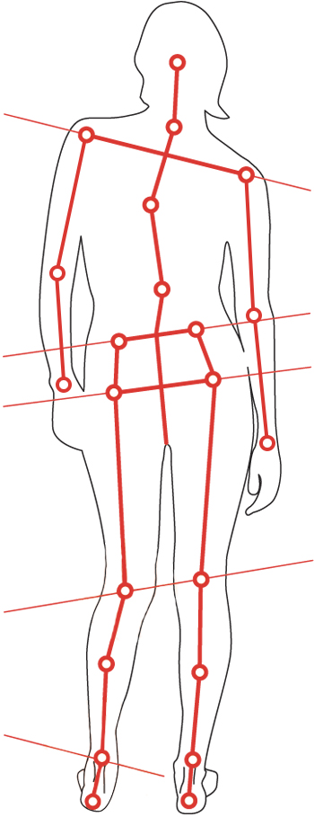
PTo detect the causes of your headaches, it is advisable to perform a OPS postural assessment.
NECK PAIN
Pathology
Cervical pain is located in the neck area. When the pain extends and radiates towards an arm, it is called cervico-brachial neuralgia. Pain can also radiate to the head through headaches.
Most often, cervical pain is favoured by inappropriate postures and movements (often called torticollis) or by arthrosis. Cervical pain can also occur after a trauma (‘whiplash’).
Poor posture of the head can lead to overstraining of the neck muscles and intervertebral discs. Poor head posture of postural origin is often caused by a maxillofacial (jaw) disorder, an oculomotor (eye) disorder, a vestibular (inner ear) disorder or even a foot disorder.
In order to detect the origin of cervical pain, it is advisable to perform an OPS postural assessment.
SHOULDER PAIN
Pathology
The origin of shoulder pain is mainly due to muscle injury. In extreme cases, it can be a fracture (of the clavicle or the humerus) or a tear (rupture of the rotator cuff in particular) or finally a dislocation (the head of the humerus coming out of its articular cavity).
Shoulder pain is mostly associated with neck pain, which is why it is essential to check the correct posture of the head.
Poor head posture of postural origin is often caused by a maxillofacial (jaw) disorder, an oculomotor (eye) disorder, a vestibular (inner ear) disorder or even a foot disorder.
In order to detect the origin of shoulder pain, it is advisable to perform an OPS postural assessment.
BACK PAIN
Pathology
Dorsalgia is a pain felt from the base of the neck to the waist (between the first and twelfth dorsal vertebrae). It is often postural in origin. Its origin can be postural descending, ascending or mixed. A poor head or feet posture can change the position of the spine and overload the intervertebral muscles and discs. A deficiency of the respiratory system can also cause a disturbance of the spine.
In order to detect the cause of back pain, it is essential to perform an OPS postural assessment combined with a detailed analysis of the mobility of the spine.
LUMBAR PAIN
Pathology
Lumbar pain is located in the lower back.
Symptoms, intensity and type of pain may vary depending on the cause. Lumbar pain can be mild or severe, periodic or chronic. It can be deep, stabbing, throbbing or pulsating. Lumbar pain can sometimes be worse in the morning and improve with movement and stretching.
Most lumbar back pain is caused by lower limb deficiencies (overpronated feet, flat feet, hollow feet, lack of cushioning, unevenness of the lower limbs, etc.). In 80% of the cases, a mixed ascending and descending postural stress is detected. The spinal column is put under tension like a cloth that twists on a vertical axis. The lower back is the central area of this tension, and it is the area that suffers the most, with a risk of wear and tear and herniated discs.
The OPS postural assessment has the advantage of analysing all possible deficiencies in posture, walking and running.
HIP PAIN
Pathology
Hip pain can occur more or less quickly. It is usually felt in the groin fold and can radiate to the knee. The origin of hip pain can be caused by muscle and joint damage.
A malposition of the foot joint can affect the hip in an ascending chain.
Inversely, a malposition of the spine can also cause a compensation of the hip in descending chain. The hip can therefore be stressed by compensation of the upper or lower stages.
The main pathologies related to hip pain are the following:
TENDINITIS OF THE ILIOPSOAS
The two muscles, iliacus and psoas, originate in the lumbar area and join to insert themselves through a tendon in a small area inside the thigh: the lesser trochanter. The iliopsoas muscle is often injured as a compensation for excessive internal rotation of the femur caused by overpronation of the foot (flat foot).
The OPS postural assessment will be all the more important because of its podiatric analysis in motion.
PYRAMIDAL (OR PIRIFORM) MUSCLE SYNDROME
The pyramidal muscle runs from the sacrum, in the lower part of the spine, through the sacroiliac joint to the top of the femoral bone or femur. The sciatic nerve passes directly below this muscle.
When the muscle becomes tense or spastic, it can cause a brief irritation of the nerve. This muscle often causes pain in the buttocks and the lumbar area. The pyramidal muscle is often injured as a compensation for the excessive internal rotation of the femur caused by overpronation of the foot (flat foot).
The OPS postural assessment will be all the more important because of its podiatric analysis in movement.
TENDINITIS OF THE GLUTEUS MEDIUS
Tendinitis of the gluteus medius is a fairly common problem. It is also called hip tendinopathy or trochanteric bursitis. The problem can occur spontaneously, or after an effort, or following the implantation of a hip prosthesis.
To compensate for the excessive internal rotation of the femur caused by overpronation of the foot, the gluteus medius muscle (external rotator of the hip) retracts to counteract the excess internal rotation of the hip. It tries to compensate for the excess amplitude of the foot in pronation (following a weakness of the supinator muscles of the foot).
The OPS postural assessment will be all the more important because of its podiatric analysis in movement.
PUBALGIA
Pubalgia is a tendinitis of one of the many abdominal muscles that end in a fibrous blade (linea alba, musculus rectus abdominis, obliquus abdominis...) or of the thigh muscles (adductor, abductor...) which are inserted on the ilio-pubic wing.
This inflammation is due to repeated and traumatic stress on the concerned tendon. The mechanism of the lesion is often linked to an asymmetry in the mobility of the 2 halves of the pelvis, mainly caused by an asymmetrical position of the 2 feet during walking or running. It is common to encounter 2 asymmetrical overpronated feet that force the pelvis to position itself in a twist that overworks the pubic symphysis.
The OPS postural assessment will be all the more important because of its global analysis in order to detect the different origins that create tension on the pubis: feet, spine...
COXARTHROSIS
Coxarthrosis (or arthrosis of the hip) is wear and tear of the hip joint caused by friction on the joint surfaces. X-rays show the destruction of the cartilage. It is common from the age of 50 onwards. Coxarthrosis can be asymptomatic and go unnoticed, or it can be very disabling and cause a lot of pain.
The malposition of the foot can cause a lack of cushioning, which can lead to excessive stress on the cartilage of the knee and hip.
Foot malposition during walking (overpronation or oversupination) can cause a poor distribution of joint pressure in the knee and hip joints, leading to abnormal cartilage wear.
The OPS postural assessment will be all the more important because of its podiatric analysis in movement.
ELBOW PAIN
Pathology
Also known as ‘tennis elbow’, epicondylitis is an inflammation that occurs near a small bony protrusion of the arm bone (humerus), just above the elbow joint on the outside of the arm. The pain mainly comes from a lesion of the tendons near the elbow. Tendons are strong bands of tissue that attach muscles to the bone.
Epicondylitis is often amplified by nocturnal cramp combining bruxism (teeth clenching) and fist clenching. This unconscious nocturnal overtiredness can cause overexertion of the epicondyle muscles. It can also be associated with a disorder of the postural mechanics characterized by an off-axis head posture with compression of the nerve roots innervating the elbow.
In order to detect the origin of elbow pain, it is advisable to carry out an OPS postural assessment.
WRIST PAIN
Pathology
The carpal tunnel syndrome manifests itself by the compression of the median nerve at the wrist. The median nerve is a large nerve that runs through the centre of the forearm and extends to the skin of the thumb, index, middle and half ring fingers.
In the wrist, the median nerve and the flexor tendons of the fingers pass through a ‘tunnel’ called the carpal tunnel. The carpal tunnel is very narrow and can be easily reduced by inflammation or its sequelae (fibrosis).
The carpal tunnel syndrome is often amplified by nocturnal cramp combining bruxism (tooth clenching) and fist clenching. This unconscious nocturnal overexertion can cause inflammation of the muscles passing through the carpal tunnel.
In order to detect the origin of the carpal tunnel syndrome, it is advisable to perform an OPS postural assessment.
KNEE PAIN
The main pathologies related to knee pain are the following:
KNEE INSTABILITY
Instability of the knee can cause misalignment of the lower limb and patella slippage. It promotes rotation of the leg and misalignment of the ankle and hip.
Knee instability is often caused by ligament hyperlaxity and muscle deficiency. Over time, it can lead to pathologies such as the patellofemoral syndrome, osteoarthritis and even patellar dislocation.
The knee is a hinge joint between the foot and the hip. A malposition of one of the two segments can cause a compensatory overload of the knee. A flat foot (overpronation) is unstable and causes compensatory problems directly at the knee.
The OPS postural assessment will be all the more important because of its podiatric analysis and its functional analysis of the hips.
PATELLOFEMORAL STRESS SYNDROME
The patellofemoral stress syndrome is also known as anterior knee pain syndrome, and has often been compared in the past to patellofemoral chondromalacia. It is frequently encountered in young athletes. This pathology is often caused by a misalignment of the lower extremity: increased femoral anteversion, tibia vara, external torsion of the tibia and pronation of the foot.
Malposition of the foot in overpronation can lead to excessive compression of the patella cartilage.
The OPS postural assessment is interesting to check the action of the foot in relation to the knee.
OSGOOD-SCHLATTER DISEASE OR ANTERIOR TIBIAL EPIPHYSITIS
Osgood-Schlatter disease is a type of osteochondritis or osteochondrosis. It is an abnormality in the growth of bone and cartilage in children. It is a group of diseases with unknown causes characterized by the interruption of the vascularization of the primary or secondary ossification nucleus of the affected bones.
Osteochondritis appears between 5 and 14 years of age depending on its location and mainly concerns individuals who are more or less athletic.
Osgood-Schlatter's disease is characterized by pain located in the anterior tibial tuberosity (upper tibia).
This osteochondrosis can be aggravated by overpronation of the foot.
The OPS postural assessment will be all the more important because of its podiatric analysis.
SINDING-LARSEN-JOHANSON DISEASE
Sinding-Larsen-Johanson disease is a type of osteochondritis or osteochondrosis. It is an abnormality in the growth of bone and cartilage in children. It is a group of diseases with unknown causes characterized by the interruption of the vascularization of the primary or secondary ossification nucleus of the affected bones.
Osteochondritis appears between 5 and 14 years of age depending on its location and mainly concerns individuals who are more or less athletic.
Sinding-Larsen-Johanson disease is characterized by pain at the tip of the patella.
This osteochondrosis can be aggravated by overpronation of the foot.
The OPS postural assessment will be all the more important because of its podiatric analysis in movement.
PATELLAR TENDINITIS (JUMPER'S KNEE)
It is a tendinitis that affects the insertion of the patellar tendon at the distal end of the patella, and, less frequently, at the anterior tuberosity of the tibia.
This tendinitis may be related to overpronation of the foot.
The OPS postural assessment will be all the more important because of its podiatric analysis in movement.
TENDINITIS OF THE GOOSE FOOT MUSCLES
Goose foot tendinitis is an inflammation of the three tendons of the muscles making up the ‘goose foot’ located in the upper-inner part of the tibia.
The inflammatory mechanism is usually caused by overpronation of the foot, which promotes torsion of the knee, putting tension on the tendons of the goose foot.
The OPS postural assessment will be all the more important because of its podiatric analysis in movement.
MAISSIAT’S BAND SYNDROME OR ‘WINDSHIELD WIPER SYNDROME’
During the effort, this Maissiat’s band (external aponeurosis of the knee) rubs abnormally against the femur like a windshield wiper. Over time, the area becomes inflamed and the person complains of external knee pain. In the majority of cases, the problem is related to a dynamic foot disorder. However, it can also be due to a pelvic problem.
The OPS postural assessment will be all the more important because of the global analysis in order to detect the origin: foot, pelvis...
GONARTHROSIS
Gonarthrosis (or arthrosis of the knee) is wear and tear of the knee joint caused by friction on the joint surfaces. X-rays show the destruction of the cartilage. It is common from the age of 50 onwards. It can be asymptomatic and go unnoticed, or it can be very disabling and cause a lot of pain.
The malposition of the foot can cause a lack of cushioning, which can lead to excessive stress on the menisci and cartilage of the knee. This malposition can cause a poor distribution of pressure in the knee joints, leading to abnormal wear of the menisci and subsequently of the cartilage.
The OPS postural assessment will be all the more important because of its podiatric analysis in movement.
GENU VALGUM
The genu valgum is an inward deformation of the lower limb. In a standing position, both legs form an X, the two knees are touching each other while the ankles are spread apart. The more pronounced the genu valgum is, the more it can interfere with walking. Also, with age, it is often a predisposing factor to gonarthrosis (arthrosis of the knee) because the pressures and forces exerted on the knee are not in the right places.
The genu valgum can be caused by an imbalance in the muscle tone of the lower limb and hyperlaxity of the internal ligaments of the knee. The foot in overpronation (flat foot) and weight excess accentuate the deformity of the knee in an X-shape.
The OPS postural assessment will be all the more important because of its dynamic analysis of the lower limbs.
GENU VARUM
The genu varum is an outward deformation of the lower limb. In standing position, both legs form an O. It is the opposite of the valgum genu (X-shaped knees). The ankles are in contact with each other, while the knees do not touch each other. The more the knee is ‘varum’, the greater the gap. The legs are called bandy or bow legs. The varum knee is one of the main factors that promote arthrosis of the knee by loading the medial compartment of the knee.
The genu varum can be caused by an imbalance between the muscle tone and flexibility of the lower limb. It can be amplified by a poor position of the feet and hips.
The OPS postural assessment will be all the more important by its dynamic analysis of the lower limbs.
WALKING WITH FEET TURNED INWARDS ‘INTOEING’
The intoeing walk is a walk with closed hips. It appears from a very young age. The feet turn inwards due to excessive internal rotation of the hip. It can lead to regular falls.
This mechanism is caused by a disturbance in the muscle tone of the hips and/or feet.
The OPS postural assessment will be all the more important because of its dynamic analysis of the lower limbs.
LEG PAIN
The main pathologies related to leg pain are the following :
TIBIAL PERIOSTITIS
Tibial periostitis manifests itself as a painful inflammatory syndrome, located along the medial two-thirds of the tibia.
The term ‘internal tibial stress syndrome’ is more specific and refers to overload injuries producing a painful inflammatory reaction along the postero-internal border of the tibia.
Tibial periostitis is related to overpronation of the foot. The muscle struggling excessively to dynamically straighten the foot inflames its bony attachment.
The OPS postural assessment will be all the more important because of its podiatric analysis in movement.
ANTERIOR TIBIAL TENDINITIS
The anterior tibial muscle extends from the bridge of the foot to the anterior part of the leg. It participates in the elevation of the forefoot in order to prepare the contact of the foot with the ground, and in the elevation of the internal arch (by supination) helping thus the posterior tibial muscle (main supinator of the foot).
Excessive pronation of the foot is the main cause of anterior tibial inflammation. Wearing a heeled shoe can also be a cause of abnormal stress since its working amplitude is more consequent.
The OPS postural assessment will be all the more important because of the analysis of the foot and shoes.
ANKLE PAIN
The main pathologies related to ankle pain are the following :
ANKLE INSTABILITY
Ankle instability is caused by joint hypermobility. It refers to every movement that occurs in a joint in response to the forces that interact with the joint when the joint should be stable under such forces.
Overpronation of the foot produces joint instability, called hypermobility, when the foot is under load. The ankle undergoes partial dislocations (subluxations) and is more prone to sprains.
The OPS postural assessment will be all the more important because of its podiatric analysis.
ANKLE SPRAIN
An ankle sprain is an injury due to stretching or tearing one or more ligaments. Ligaments are elastics stretched from one bone to another and allowing the joints to remain stable in the event of extreme movements. Ninety percent of ankle sprains correspond to damage to the external collateral ligament between the tibia, the talus (astragalus) and the calcaneus (heel bone).
Following a first sprain, statistics show that a recurrence occurs in 50% of cases. It is therefore important to prevent this phenomenon by carrying out adequate proprioceptive and muscular rehabilitation.
A flat foot (overpronation) is unstable when it is caused by muscle weakness. Its malposition can therefore disturb the stability of the foot. The hollow foot (oversupination) is the opposite of the flat foot, which is too rigid. Its lack of mobility makes the ligaments vulnerable to the slightest variation of the ground.
The OPS postural assessment will be all the more important because of its podiatric analysis.
FOOT PAIN
The main pathologies related to foot pain are the following :
FLAT FOOT
The flat foot is characterised by a sagging of the arch of the foot with a decrease in the height of the inner arch of the foot. The phenomenon is usually accompanied by excessive pronation (overpronation).
Some parents are concerned about their child's flat feet. First of all, it is important to know that at birth, the foot is chubby with excess fat which is reduced in the first two years of life. From this age onwards, the foot will start to take shape according to its curvature. It is also important to know that the arch of the foot begins to take form through the activity of the foot muscles during walking. A child completes the muscular and proprioceptive development of his feet at the age of 8 years. An assessment is therefore necessary to visualise the development of a child's feet. The foot is therefore flat at its first steps and its deformity is progressively reduced until the age of 8 years, provided that it has a correct muscular development.
The congenital flat foot is said to be caused by muscular insufficiency and ligamentous distension of the foot which would not allow the arch to position itself normally.
An acquired flat foot is the result of a deformity of the foot in which the position of the bones in relation to each other has been altered. This modification can be caused by many factors which disrupt muscle and ligament function (footwear, trauma, etc.).
The OPS postural assessment will be all the more important because of the podiatric analysis.
HOLLOW FOOT
The hollow foot (oversupination) is characterised by the exaggerated accentuation of the arch. Specifically, it means an increase in height of the arches of the foot and is regularly accompanied by claw toes. The hollow foot is often rigid and unstable. It is therefore more easily subject to external ankle sprains.
The OPS postural assessment will be all the more important because of the podiatric analysis.
FOOT INSTABILITY
Instability of the foot is caused by hypermobility of the joints. It refers to every movement that occurs in a joint in response to the forces that interact with the joint when it should be stable under such forces.
Overpronation of the foot produces joint instability, called hypermobility when the foot is under load. This hypermobility is the cause of certain problems such as metatarsalgia, subluxation of the metatarsophalangeal joint and neuromas.
When the foot is in pronation during propulsion, the bone locking mechanism of the tarsus becomes less effective. The tarsus becomes unstable, the muscles in the support phase are unable to effectively stabilise the distal joints. No distal bones can be stabilised if the proximal bones are unstable. When the heel detaches from the ground, the forces supported by the forefoot cause abnormal movements of the forefoot bones towards each other, as well as a decrease in bone stability. Partial dislocations (subluxations) occur in the joint.
The OPS postural assessment will be all the more important because of its podiatric analysis.
METATARSALGY
Metatarsalgia is a non-specific term that defines pain in the metatarsals, generally in the metatarsophalangeal joint.
Chronic metatarsalgia is mostly caused by microtraumas produced by overpronation of the foot during propulsion. As the foot cannot be locked, its deviated and hypermobile joints stretch the ligament and capsular tissues, causing inflammation. Overpronation of the foot can also cause abnormal shearing on the heads of the metatarsals, which can lead to pain.
An anterior fall of the trunk can also cause an overload of weight on the front of the foot. Anterior head posture can be caused by maxillofacial imbalance (jaws), oculomotor (eyes) or vestibular (inner ear) disorders.
The OPS postural assessment will be all the more important because of its global vision.
NEUROMA OR MORTON DISEASE
Intermetatarsal neuroma, also known as Morton's syndrome, is a pain that develops in the region of the bifurcation of the neurovascular bundle passing between the toes.
The pain generally occurs in the third intermetatarsal space, more rarely in the second one and, exceptionally, in the first and fourth one. This pain is of the ‘electric discharge’ type, often forcing the patient to take off his shoes in order to calm the symptoms. It can be awakened by punctiform pressure on the plantar surface and is frequently accompanied by irradiation towards the adjacent toes, which are the seat of paraesthesia.
Morphostatic disorders of the foot (overpronation) and the wearing of narrow shoes with high heels are predisposing factors. Likewise, heavy sporting activities can facilitate the occurrence of this pathology due to the micro-traumatic stresses they generate.
The OPS postural assessment will be all the more important because of its podiatric analysis.
HALLUX VALGUS
The hallux valgus of the foot is a deviation of the big toe towards the outside of the foot. Its deformation mechanism is mainly caused by prolonged excessive pronation of the foot. The unlocked medio-tarsal joint induces hypermobility of the first radius during propulsion. The unstable first ray is forced into dorsiflexion and inversion, causing subluxation of the first metatarsophalangeal joint. The deformation is progressive and influenced by the degree of pronation.
The OPS postural assessment will be all the more important because of its podiatric analysis.
CLAW DEFORMITY
This is a deformity of a toe at the proximal and/or distal interphalangeal joint. It can be isolated or associated with hallux valgus.
It is favoured by the wearing of shoes with too narrow toes and high heels. A malpositioned foot (overpronated or oversupinated) can lead to instability of the foot and promote the formation of toe claws. This prolonged claw formation can become rigid over time and end up in a claw position.
An anterior fall of the trunk can also cause a weight overload on the front of the foot and lead to claw formation of the toes to improve the stability of the body. An anterior head posture, responsible for anteriority of the trunk, can be caused by a maxillofacial imbalance (jaws), an oculomotor disorder (eyes) or a vestibular disorder (inner ear).
The OPS postural assessment will be all the more important because of its global analysis.
TENDINOPATHIES OF THE ACHILLES TENDON
The Achilles tendon is the tendon of the body which is subject to the greatest stress, up to several hundred kilos of traction for a minimum size of about 1 cm². It is better to speak of tendinopathy than tendinitis, given the anatomopathological diversity.
Tendinopathies of the Achilles tendon are often related to overpronation of the foot.
The OPS postural assessment will be all the more important because of its podiatric analysis.
SEVER’S DISEASE
Sever's disease is a type of osteochondritis or osteochondrosis. It is an abnormality in the growth of bone and cartilage in children. It is a group of diseases of unknown causes characterised by the interruption of the vascularisation of the primary or secondary ossification nucleus of the affected bones.
Osteochondritis appears between 5 and 14 years of age depending on its location and mainly concerns individuals who are more or less athletic.
Sever's disease is characterised by pain in the calcaneus (back of the heel).
Sever's disease is often aggravated by overpronation of the foot. Excessive pronation is accompanied by an internal rotation of the tibia which tends to pull the Achilles tendon inwards with a quick whiplash. This can therefore lead to injury of the cartilage insertion with which the tendon attaches itself.
The OPS postural assessment will be all the more important because of its podiatric analysis.
TENDINOPATHY OF THE HIND LEG
Tendinopathy of the hind leg is quite frequent and is clearly favoured by overpronation of the foot. It may be an insertion tendinopathy on the tarsal scaphoid or a tenosynovitis behind the internal malleolus. Dislocation of this tendon is rare. However, degenerative tendinopathy with progressive stretching and then partial and complete rupture of the tendon is more frequent.
The OPS postural assessment will be all the more important because of its podiatric analysis.
PLANTAR APONEUROSITIS
The plantar aponeurosis or fascia is a fibromuscular structure that is part of the sural-Achilles-plantar system. It is poorly vascularised and more elastic. It is called upon in the event of impulse movements and sagging feet.
It is most often at the posteromedial level, at its insertion on the calcaneum, that the plantar aponeurosis presents a painful inflammation. This pain can radiate towards the big toe. It is typically present when the foot is first placed on the ground in the morning, after resting at night. There is an palpatory pain, which is revived at dorsiflexion of the big toe. X-rays will usually be normal in young sports patients, but will show a ‘calcaneal spur’ in the older patient. This spur is the result of traction of the fascia on the calcaneus, but is not painful on its own.
Plantar fascia also appears to be related to overpronation. In fact, the prolonged eversion of the calcaneus during the intermediate support phase leads to a stretching of the plantar aponeurosis. If the foot does not return to supination, the tension of the plantar aponeurosis increases at the moment the heel is detached from the ground, when the weight of the body is concentrated on the forefoot and when the triceps by its contraction exerts a traction on the calcaneus. Excessive pronation coupled with these factors during running causes an enormous increase in tension which results in microtraumas of the plantar fascia due to repetitive overloading and stress build-up.
LENOIR’S SPUR
Lenoir's spur is what is more commonly known as the calcaneal spur. It is an abnormal bone excrescence (pointing towards the forefoot), visible on X-rays, named in honour of Mr Lenoir (who made its discovery). Calcaneal spur forms where the plantar fascia meets the heel bone (calcaneus). It is not a disease in itself, but a consequence of chronic plantar fasciitis (also known as plantar aponeurositis).
The OPS postural assessment will be all the more important because of its podiatric analysis.
STRESS FRACTURE
Bone tissue is able to repair its structure in response to stress. The normal remodelling process requires a balance between bone resorption and bone replacement. Microfractures therefore occur when the resorption process is faster than the replacement process. This imbalance is caused by the continuous application of repetitive stress. The calcaneus and metatarsals are the most common locations of stress fractures in the athlete.
The aetiology of stress fractures is multifactorial :
- Training intensity,
- Dietetics,
- Overpronation of the foot: this overloads the soleus muscle which can tire the posteromedial part of the tibia and cause a stress fracture. In addition, this overpronation is likely to lead to a stress fracture of the calcaneus due to the lack of shock absorption caused by the absence of ‘spring action’ of the foot.
The OPS postural assessment will be all the more important because of its podiatric analysis.
ALGODYSTROPHY OF THE FOOT
Algodystrophy or algoneurodystrophy, also known as ‘complex regional pain syndrome’, manifests itself by tingling, burning, intense pain and even stiffness. Its cause is often difficult to find, and in 20% of cases it remains mysterious. Algodystrophy occurs in the joints and peri-articular areas. It is essential to combat joint stiffness as early as possible in order to avoid complete fixation of the affected joint. Active rehabilitation is essential.
The OPS postural assessment will enable to analyse the level of joint damage (stiffness) and to visualise the body's compensation for this pathology.
DIABETIC FOOT (NEUROPATHY)
Neuropathy is one of the most recurrent complications of diabetes. It first affects the feet and then the hands. It is described as a loss of sensation, or numbness or tingling in the toes. Symptoms usually appear progressively with diffuse and intermittent pain at the beginning and may intensify over time. Symptoms come and go, but eventually become more frequent. Over a long period of time, a person may experience a loss of sensation to the point where they cannot feel the tightness of their shoes, or don’t even know if the bath water is hot or cold, or if they have injured themselves.
Symptoms of muscular weakness and progressive sagging of the arches of the foot also appear. Diabetic neuropathy is the main cause of foot ulcerations and infections and, in extreme cases, amputation.
It is essential to check that the foot does not suffer from a pressure disorder that could lead to ulceration.
The OPS postural assessment will be all the more important because of its podiatric analysis.